
Superior Diagnostic Services
Our Laboratory resources include:
Please contact Elizabeth Weiler at 617-358-9740 or e-mail at contact@skinpathlab.com to arrange for service or to ask any questions you may have.
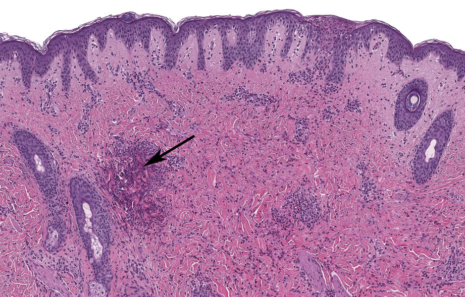
Focal erosion, mild papillary dermal edema, and mild to moderate perivascular and interstitial lymphohistiocytic infiltrate with a flame figure (arrow) in an arthropod bite reaction.
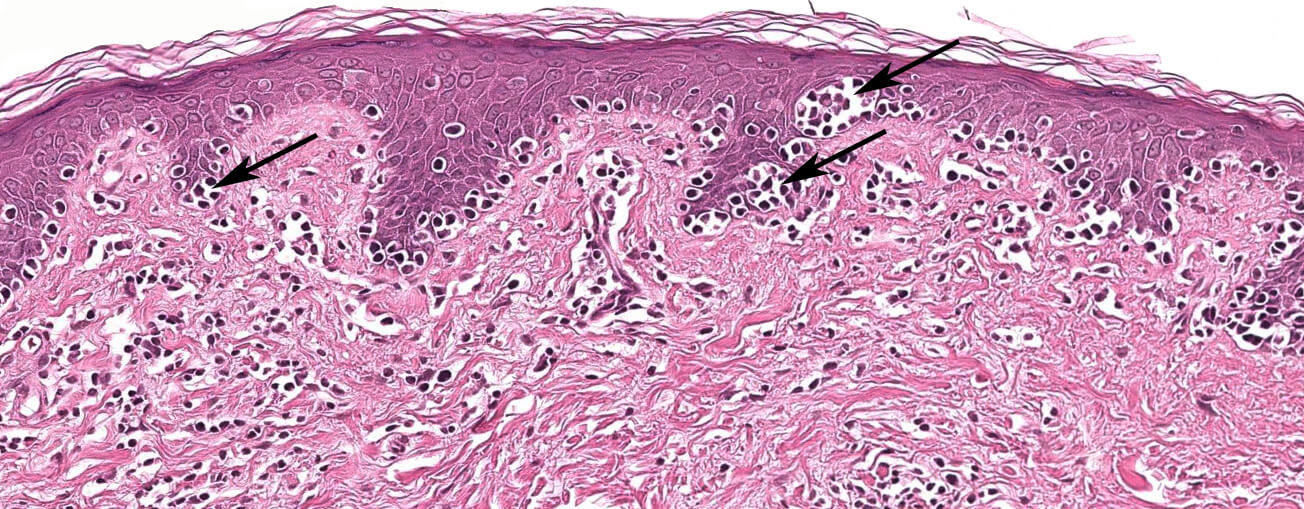
Predominantly intra-epidermal atypical lymphocytic infiltrate with Pautrier microabscesses (arrow) in cutaneous T-cell lymphoma.

Multiple neutrophilic microabscesses (arrow) located at the tips of the dermal papillae in dermatitis herpetiformis.
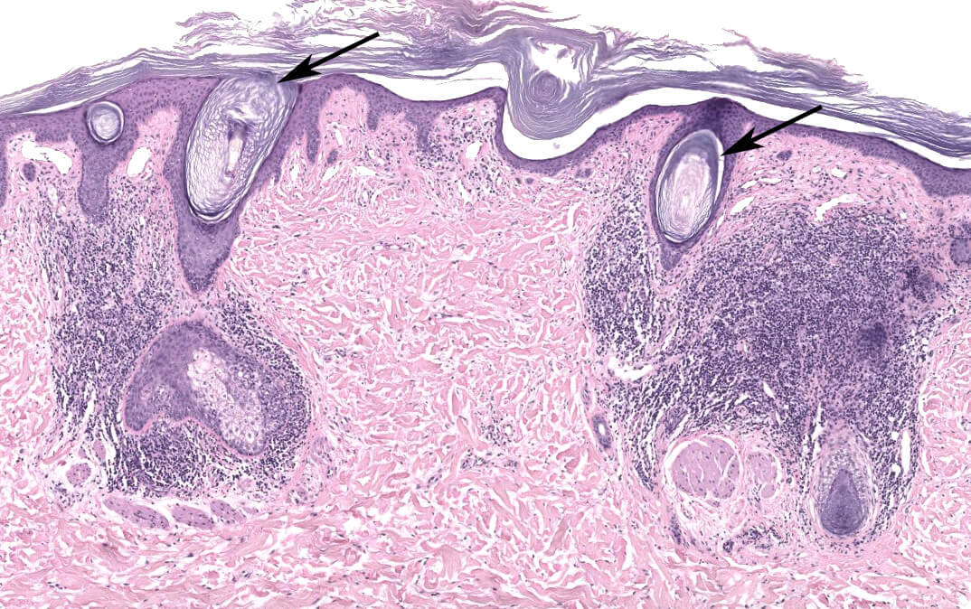
Hyperkeratosis, follicular plugging (arrow), epidermal atrophy, and moderate to dense perivascular and perifollicular lymphocytic infiltrate in discoid lupus erythematosus.
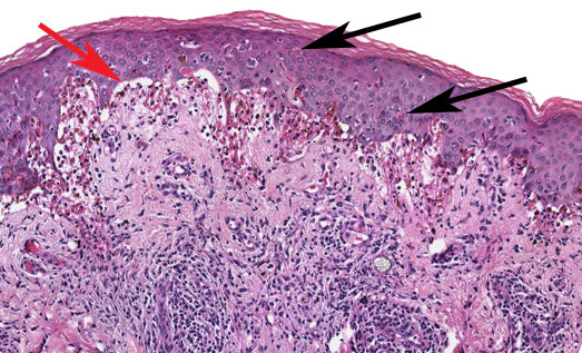
Interface dermatitis with numerous individually necrotic keratinocytes (black arrow), basal cell layer vacuolization (red arrow), papillary dermal edema, and moderate perivascular lymphohistiocytic infiltrate in erythema multiforme.
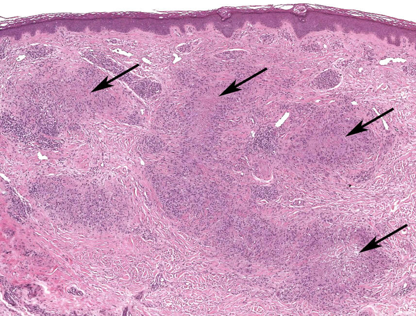
Several palisaded histiocytic granulomas (arrow) containing mucin and a perivascular lymphohistiocytic infiltrate in granuloma annulare.
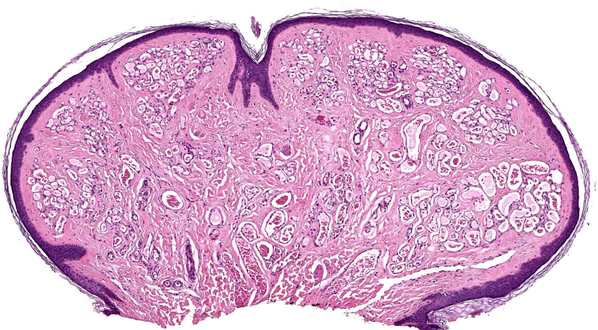
Epidermal collarette surrounding a well-circumscribed and lobular proliferation of small vessels within the papillary dermis in a hemangioma.
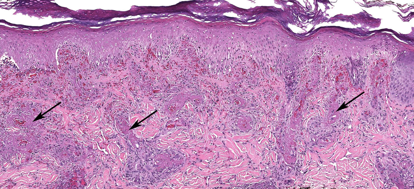
Fibrinoid necrosis of vessel walls (arrow) with extravasation of erythrocytes in leukocytoclastic vasculitis.
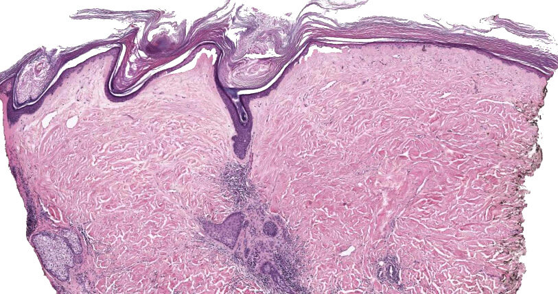
Hyperkeratosis with follicular plugging, atrophy of epidermis and homogenization of the collagen in the papillary dermis (arrow) in lichen sclerosus (et atrophicus).
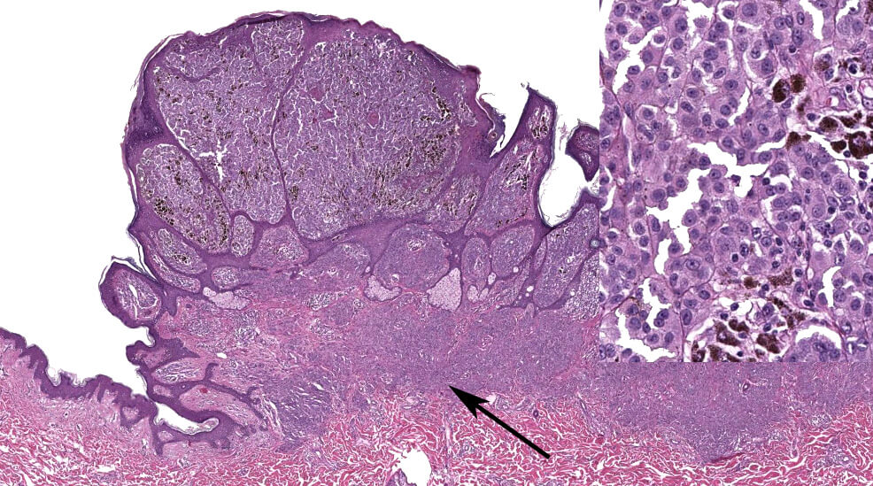
Polypoidal lesion with markedly atypical melanocytes (see inset) consistent with malignant melanoma arising from an underlying nevus (arrow).

Nested and lentiginous intra-epidermal atypical melanocytic proliferation in malignant melanoma in situ.
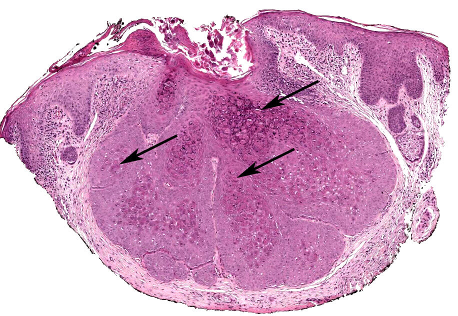
Crateriform, endophytic lobular epidermal hyperplasia, with intracytoplasmic purple-red, oval inclusions (molluscum bodies) in the supra basal keratinocytes (arrow) in molluscum contagiosum.
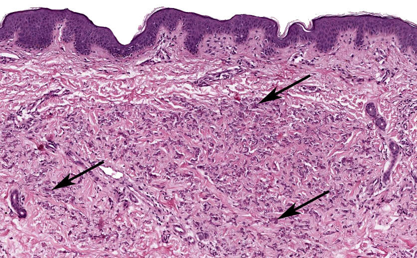
Clumped, fragmented and curled basophilic elastic fibers (arrow) in the middle and lower third of dermis in pseudoxanthoma elasticum.
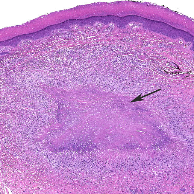
Mid to deep dermal palisaded histiocytes surrounding a central area of irregularly shaped, eosinophilic necrobiosis (arrow) in a rheumatoid nodule.
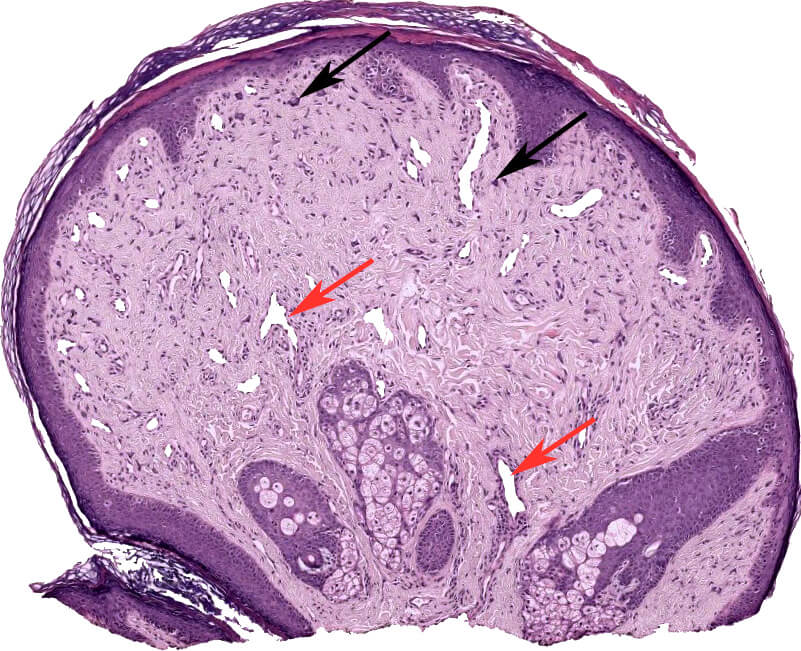
Polypoid lesion containing stellate and multinucleated fibroblasts (black arrow) and increased numbers of telangiectatic vessels (red arrow) in fibrous papule.
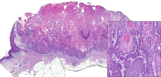
Hyperkeratosis, epithelial hyperplasia, and irregular islands of squamous cells with central keratinization with horn pearl formation in the dermis (see inset) in a squamous cell carcinoma.

Hyperkeratosis, irregular epidermal hyperplasia, with full thickness keratinocytic atypia with hyperchromatic nuclei and pale cytoplasm in squamous cell carcinoma in situ.
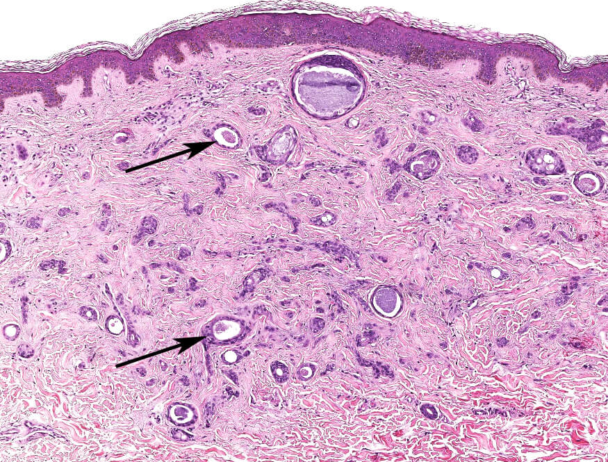
Short interconnecting epithelial strands with occasional ectatic ducts or microcysts forming a paisley tie or tadpole pattern (arrow) and set in a fibrotic stroma within the superficial reticular dermis in a syringoma.
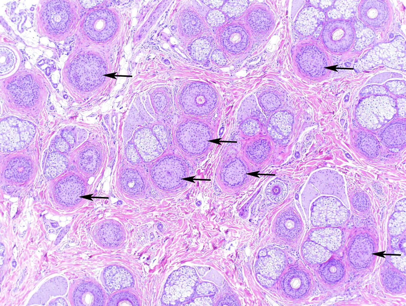
Alopecia areata with a shift into catagen and telogen (black arrow).
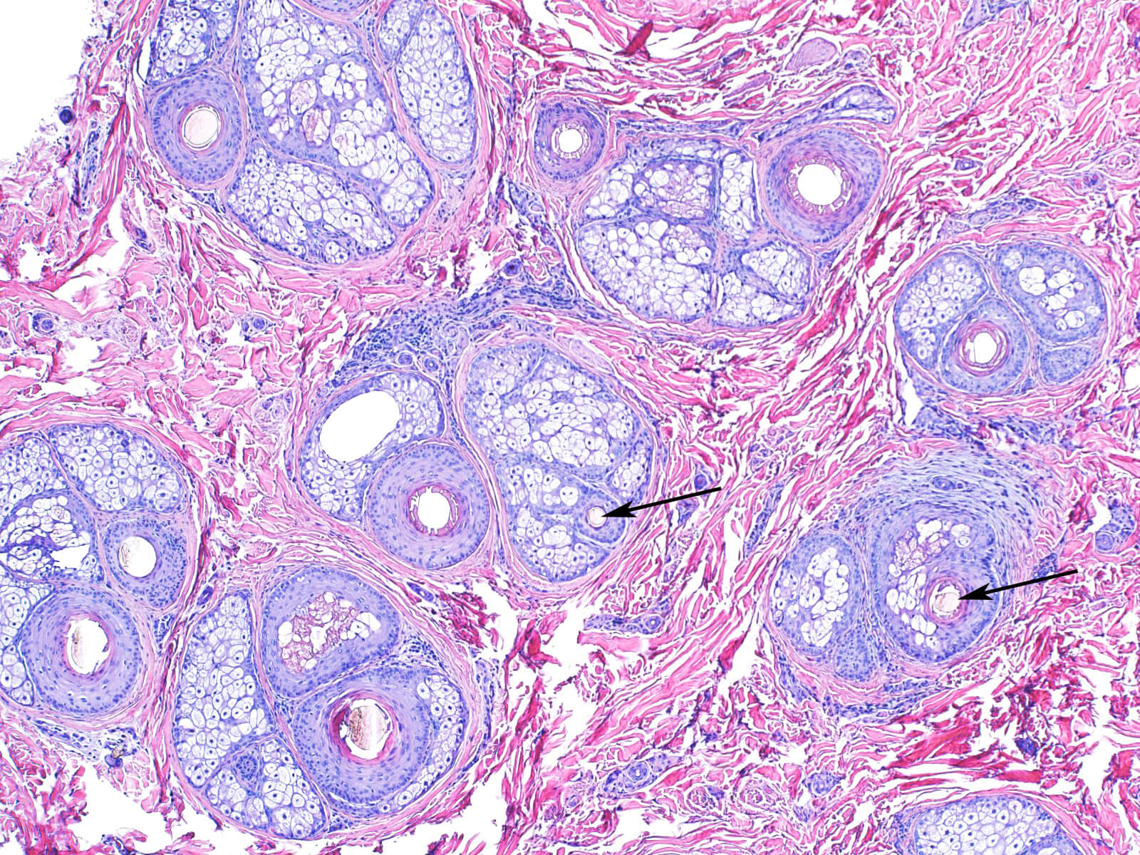
Female pattern hair loss with an increase in small hair shafts (black arrow).
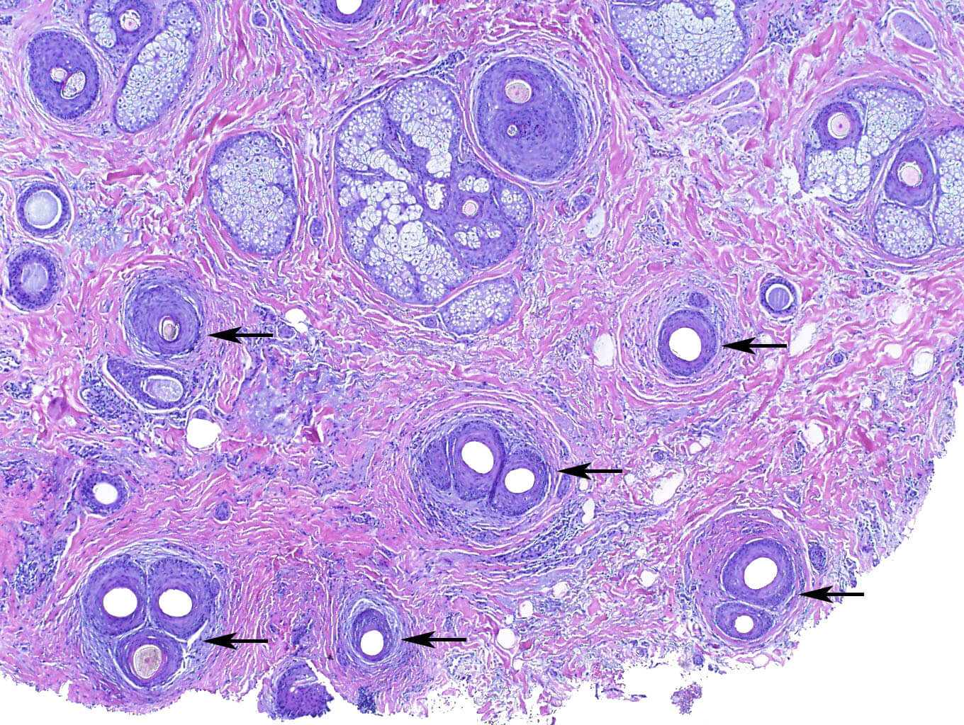
Lymphocytic scarring alopecia focally affecting several follicular units (black arrow).
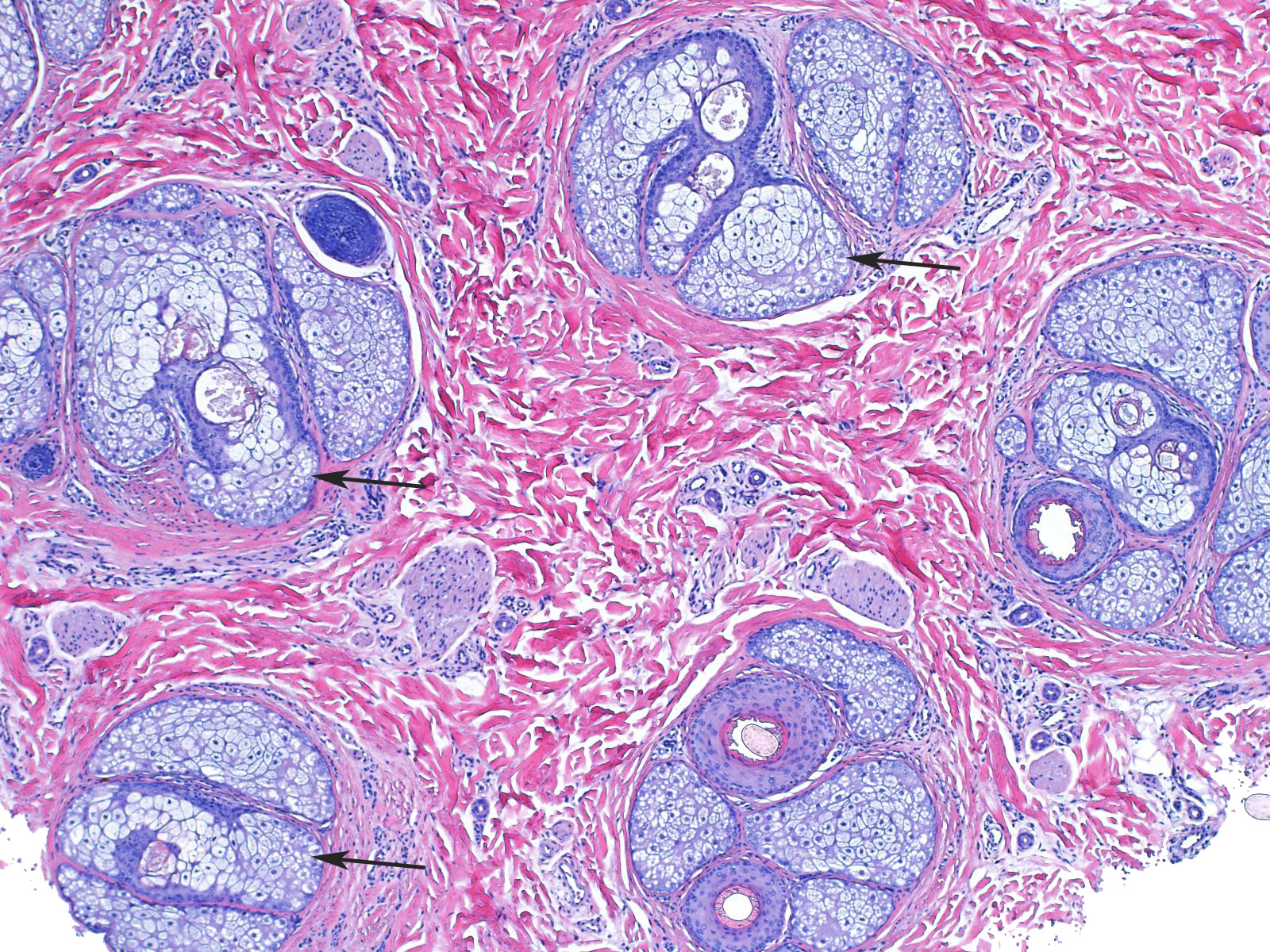
Traction alopecia looks deceptively normal but hair follicles are lacking, while sebaceous glands are retained (black arrow).


