Research
Skin Pathology Laboratory offers evaluation of skin samples utilizing all the above-described techniques. We have performed evaluations of several studies including multicenter studies. We are involved in studies of photoaging, disorders of pigmentation including vitiligo, and melasma, psoriasis, wound healing, melanoma, small nerve fiber associated atrophy, laser resurfacing and many others. Please see some selected references (Reference). We have the capability to process multiple tissues from one or many patients to avoid staining variability.
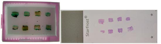
Block embedded with 8 skin samples (left panel) and H&E stained section (right panel).
New diagnostic tool for patient care and research
Most recently we have developed a technique to evaluate small nerve fibers. Cutaneous innervation plays a pathophysiologic role in disease states of the skin as well as being targeted in diseases involving other organ systems. In this regard, Skin Path Lab now offers the ability to generate intraepidermal nerve fiber counts using conventional two-dimensional analysis in vertically-oriented skin sections (see Figure 1, A), such as is currently used in the detection of small fiber neuropathies. Moreover, we have developed a new technique to visualize nerve fibers in horizontal secions, affording three-dimensional assessment of nerve fiber quantity and distribution in skin biopsies. Depending on the antibody used, we can visualize all nerves in the skin (PGP9.5, see Figure 1, B), as well as specific subsets, including p75 NGFRc-positive nerves in the dermis, autonomic (tyrosine hydroxylase—see Figure 1, C; vasoactive intestinal peptide) and sensory nerves (CGRP and substance P receptor). This newer technique preserves the architecture of the nerves in relation to the surrounding epidermal and dermal structures including at the dermal-epidermal juction, overcomes sampling bias inherent to the traditional 2D method that can lead to false negative or positive test results, and will be used to understand and diagnosis non-skin and skin diseases.
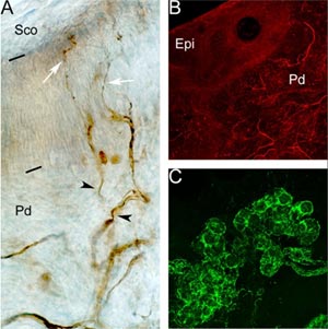
Figures. (A) Two dimensional intra-epidermal nerve fiber staining using PGP9.5 antibody , 400x. Black bars delineate the epidermis (Epi) from the overlying stratum corneum (Sco) and underlying papillary dermis (Pd). Slender intraepidermal nerve branches (white arrows) can be seen arising from the immediately subjacent subepidermal nerve plexus (black arrowheads). (B,C) Three dimensional imaging, in a 200 micron thick transverse skin section. (B) Detection of intraepidermal and papillary dermal nerve fibers using PGP9.5 antibody, 100x. (C) Autonomic adrenergic innervation of sweat glands (tyrosine hydroxylase, green) , 200x.