Plastic Sectioning and Electron Microscopy
Our Laboratory provides complete service from preparation of one-micron thick sections to ultra thin sections and preparation of electron micrographs. These procedures have been used not only in diagnosis but in research applications (Ref.)
Plastic sectioning:
Allows to examine the tissue detail used in diagnosis of lymphoid malignancies and for teaching & research purposes.

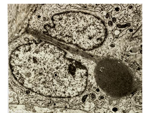
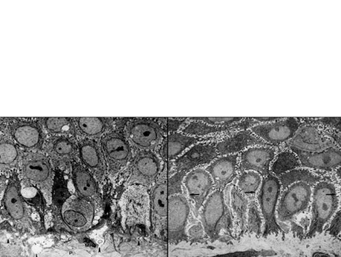
Electronmicroscopy: